Radiology
Radiology is a branch of medical science in which various forms of radiant energy are used to diagnose and treat disorders and diseases. For nearly 80 years, radiology was based primarily on the use of X rays. Since the 1970s, however, several new imaging techniques have been developed. Some, like computed tomography, makes use of X rays along with other technology, such as computer technology. Others, like ultrasound and magnetic resonance imaging, use forms of radiant energy other than X rays.
Radiant energy
The term radiant energy refers to any form of electromagnetic energy, such as cosmic rays, gamma rays, X rays, infrared radiation, visible light, ultraviolet radiation, radar, radio waves, and microwaves. These forms of energy are classified together because they all travel by means of waves. They differ from each other only in their frequencies (the number of times per second that waves vibrate) and wavelengths (the distance between two peaks in any wave).
Words to Know
Angiography: Imaging of a blood vessel by injecting a radiopaque substance in the bloodstream and exposing the body to X rays.
Computerized axial tomography (CAT): A body imaging technique in which X-ray photographs taken from a number of angles are combined by means of a computer program.
Diagnosis: Identification of a disease or disorder.
Electromagnetic radiation: Radiation that transmits energy through the interaction of electricity and magnetism.
Imaging: The process by which a "picture" is taken of the interior of a body.
Myelography: Imaging of the spinal code by radiologic techniques.
Positron emission tomography (PET): A radiologic imaging technique that makes use of photographs produced by radiation given off by radioactive materials injected into a person's body.
Radiant energy: Any form of electromagnetic energy.
Radiation therapy: The use of X rays or other radioactive substances to treat disease.
Radiopaque: Any substance through which X rays cannot pass.
Ultrasound: A form of energy that consists of waves traveling with frequencies higher than can be heard by humans; also, a technique for imaging the human body and other objects using ultrasound energy.
X rays: Electromagnetic radiation of a wavelength just shorter than ultraviolet radiation but longer than gamma rays that can penetrate solids.
Various forms of radiant energy interact with matter in different ways. For example, visible light does not pass through most forms of matter. If you hold a sheet of paper between yourself and a friend, you will not be able to see your friend. Light waves from the friend are not able to pass through the paper.
Forms of radiant energy with higher frequencies than visible light are able to penetrate matter better than does visible light. For example, if you were to place a sheet of paper between yourself and an X-ray machine, X rays would be able to pass through the paper and to strike your body.
X rays for diagnosis
The ability of X rays to pass through matter makes them useful as a diagnostic tool to identify a disease or disorder. As an example, suppose that a doctor believes that a child has broken a bone in her arm. In order to confirm this diagnosis, the doctor may take an X ray of the child's arm. In this process, the child's arm is placed beneath a machine that emits X rays. Those X rays pass through flesh in the arm without being stopped. But the X rays are not able to pass through bone as easily. A photographic plate placed beneath the child's arm "takes a picture" of X rays that have passed through the arm. Fleshy parts of the arm show up as exposed areas, while bone shows up as unexposed areas. A doctor can look at the photograph produced and determine whether the bone is solid or has been broken. Making pictures of the interior of a person's body by a process such as this is known as imaging.
Over the years, radiologists have developed more sophisticated ways of using X rays for diagnosis. For example, regions of the body in which tissue is more dense than in other regions can be detected by X-ray imaging. The presence of such dense spots may indicate the presence of a tumor or some other abnormal structure.
Radiologists also make use of substances through which X rays cannot pass, substances that are called radiopaque. Suppose that a radiopaque substance is injected into a person's bloodstream and an X ray made of the person's arm. This process is known as angiography. The radiopaque substance in the bloodstream will show up on the X-ray photograph and allow a doctor to determine the presence of abnormalities in veins, arteries, or other parts of the circulatory system.
Another form of angiography is called myelography. In this process, a radiopaque substance is injected in the membrane covering the spine, and an X-ray photograph is taken. The resulting image can be used to diagnose problems with the spine.
Computers and radiology
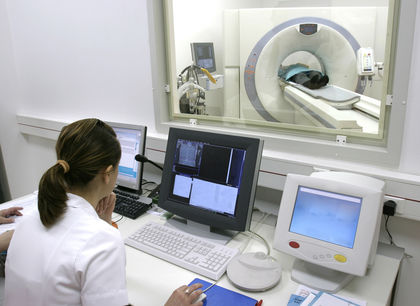
In recent decades, radiologists have developed a variety of techniques in which the powers of X rays and computers have been brought together. The earliest of these techniques was computerized axial tomography (CAT). In computerized axial tomography, an X-ray machine is rotated around a person's body. Pictures are taken of some specific part of the body from many different angles. Those pictures are then put together by a computer to provide a three-dimensional image of the body part being studied.
A variation of the CAT technique is known as positron emission tomography (PET). In this technique, a radioactive material is injected into a person's body. That radioactive material emits positrons (positive electrons) and gamma rays. A scanner "reads" the gamma rays in much the same ways that X rays are scanned in a CAT machine. However, the specific radioactive material used in the process can be chosen to produce much finer images than are available with a CAT scan. Another variation of the PET process is called single photon emission computerized tomography (SPECT).
One of the first techniques used in radiology not based on X rays was ultrasound. Ultrasound is a form of energy that consists of waves traveling with frequencies higher than can be heard by humans. Ultrasound has some of the same abilities to pass through human tissue as do X rays. One of the first uses of ultrasound was to detect defects in metallic structures. Later, it became a common and powerful tool for imaging a fetus while it is still in the uterus (womb). In this procedure, a sound transmitter is used to send waves into the pregnant woman's body from various angles. As these waves bounce back off the uterus and the fetus, they are recorded both on a television screen and in a photograph. With this technology, a physician can recognize problems that may exist within the fetus or the pregnant woman's uterus.
Therapeutic applications
Radiological techniques can also be used for therapeutic purposes, methods used to treat diseases and disorders. The use of radiology for therapy depends on the fact that X rays kill living cells. Under normal circumstances, this fact provides a good reason for people to avoid coming into contact with X rays. The destruction of healthy cells by X rays is, in fact, one of the ways in which cancers may develop.
This same fact, however, provides the basis for treating cancer. Cancer is a disease characterized by the rapid, out-of-control growth of cells. Suppose that a person has been diagnosed with cancer of the spleen, for example. That diagnosis means that cells in the spleen have begun to grow much more rapidly than normal. It follows that one way to treat this condition is to bombard the spleen with X rays. Since the cancer cells are the cells growing most rapidly, they are most likely to be the cells killed by the X rays. The fact that healthy cells are also killed in this process is shown by the side-effects of radiation therapy: loss of hair, nausea, loss of weight, among others. In fact, the success of radiation therapy depends to some extent on the physician's ability to focus the cell-killing X rays on cancer cells and to protect healthy cells from those same X rays.
[ See also X rays ]