Embryo and embryonic development
The term embryo applies to the earliest form of life, produced when an egg (female reproductive cell) is fertilized by a sperm (male reproductive cell; semen). The fertilized egg is called a zygote. Shortly after fertilization, the zygote begins to grow and develop. It divides to form two cells, then four, then eight, and so on. As the zygote and its daughter cells divide, they start to become specialized, meaning they begin to take on characteristic structures and functions that will be needed in the adult plant or animal.
An embryo is a living organism, like a full-grown rose bush, frog, or human. It has the same needs—food, oxygen, warmth, and protection—that the adult organism has. These needs are provided for in a variety of ways by different kinds of organisms.
Embryology
The study of changes that take place in the embryo is known as embryology. As one might imagine, the subject of embryology has fascinated humans since the dawn of time. Every culture has had its own theories and beliefs as to how the young of any species are created and born. The earliest formal writings on embryology can be traced to about 1416 B.C. in India. A document in Sanskrit (an ancient Indian language) describes the origin of the embryo being the union of the blood from the mother and semen from the father. Although this is not completly accurate, the document goes on to describe various stages of embryo development.
Words to Know
Differentiation: The process by which cells mature into specialized cell types, such as blood cells, muscle cells, brain cells, and sex cells.
Ectoderm: The outer layer or cells in the multilayered embryo.
Endoderm: The innermost wall of a multilayered embryo.
Fetus: In the higher vertebrates, the complex stage of development that follows the completion of the embryonic stage until hatching or birth.
Mesoderm: The central layer of cells in an embryo covered by three walls.
Ultrasonography: A process used to obtain "pictures" of the developing embryo using ultrasound.
Zygote: A fertilized egg.
Our modern understanding of changes that take place within the embryo can be traced to the rise of the cell theory in about 1838. Scientists finally discovered the process by which sperm cells from a male and egg cells from a female combine to form a zygote. Studies by the Austrian monk Gregor Mendel (1822–1884) opened a way to explain how genetic characteristics were transmitted from one generation to the next. Finally, in 1953, the discovery of the molecular structure of DNA (deoxyribonucleic acid) by the American biologist James Watson (1928– ) and the English chemist Francis Crick (1916– ) provided a chemical explanation of changes that take place during fertilization and development.
Embryonic development
The term embryonic development refers to changes that take place as an embryo matures. Those changes differ from plants to animals and from species to species. The discussion that follows focuses on embryonic development in humans.
The zygote forms in one of the mother's fallopian tubes, the tubes that connect the ovaries with the uterus. It then travels to the uterus, where it becomes affixed to the uterine lining. Along the way, the zygote divides a number of times. By the time it reaches the uterus, it consists of about 100 cells and is called an embryoblast.
The exact day on which the embryoblast implants on the uterine wall varies, but is usually about the sixth day after fertilization. By the end of the first week, a protective sac, the amniotic cavity, begins to form around the embryoblast. Changes now begin to take place at a rapid rate.
During week two of embryonic development, embryonic cells have begun the process of differentiation. The identical cells formed by the early divisions of the zygote are beginning to take on the different characteristic of muscle, blood, nerve, bone, and other kinds of cells. The embryo has burrowed deep into the uterine wall and is visible as a bump on the inner uterine surface. This position permits the embryo to receive oxygen and nutrients from the mother's blood and to excrete waste products into her bloodstream.
Miscarriages are not uncommon at this stage of pregnancy. The mother's immune system may react to cells from the embryo that it classifies as "foreign" and will begin to attack those cells. The embryo may die and be expelled.
During week three the embryo grows to a length of about 0.08 inches (2 millimeters) long and has become pear-shaped with a rounded head and a tapered tail end. Three distinct types of cells can be distinguished. Ectoderm cells will form the embryo's skin; mesoderm cells its bones, muscles, and organs; and endoderm cells its digestive tract.
Blood vessels have begun to form and, by day 20, the embryo has developed its own arteries and veins. Cells begin to collect along the embryo's back in a formation known as the neural tube, a structure that will eventually develop into the brain and spinal cord.
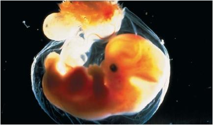
During the fourth week, the embryo becomes C-shaped with an enlarged forebrain and a visible tail. Eye stalks and ear pits appear. Upper and lower limb buds are observable. Lung, liver, pancreatic, and gall bladder buds emerge. The umbilical cord and early facial areas also form. By the end of this week, the embryo is comprised of millions of cells and is about 0.12 to 0.16 inches (3 to 4 millimeters) long. To the naked eye, the embryo looks like a small oval.
Extensive neural (nerve) and cardiac (heart) development takes place this week. Early bone formations, that will later be the vertebrae, appear along the neural tube. Nerves, muscle, and connective tissues emerge around the primitive bone formations.
By the end of fifth week, the embryo is almost 0.5 inch (about 7 to 9 millimeters) long and has all of its internal organs. The external ears emerge, and upper limb buds extend to form paddlelike hands. The mouth, stomach, and urinary bladder are present. Nose pits and eye lenses are visible. A few days after upper limb bud extension, the lower limb buds evolve further. Much more brain development occurs at this time, and the head enlarges, causing it to bend forward and appear large compared to the body. The umbilical cord becomes more clearly defined.
During the sixth week, the trunk straightens and upper limb development continues. The neck, elbows, and wrists form. Mammary and pituitary gland buds appear. Bone, cartilage, and muscles become defined around the spinal cord and in the embryonic chest. Early in this week, tooth buds appear. These buds will become the "baby" teeth that are lost in childhood. Rib cells line up horizontally along the trunk sides, and skin
layers that will hold sweat glands develop. The regions of the brain that will become the cerebral hemispheres are very prominent at this time. The embryo appears more human by this point. It is about 0.44 to 0.56 inch (11 to 14 millimeters) long, and its heart is beating at the rate of 140 to 150 beats per minute.
During the seventh week, future fingers and thumbs are clearly visible on the hands. The torso lengthens, the tail begins to disappear, and the primitive organs continue to evolve. The heart has become divided into chambers. The cornea of the eye is also present. By the end of this week, the embryo is about 0.8 inch (20 millimeters) long and about the size of a quarter.
During the eighth week, remarkable development occurs. Nerve cells in the brain form at a rate of about 100,000 a minute. The top of the head becomes more rounded and erect. Between day 52 and day 56, the fanshaped toes go from being webbed to separated. The fingers are entirely distinct. The eyelids close over the eyes and become fused shut until about the twenty-sixth week. External genital (sex organ) differences begin to develop. All appearances of the tail are gone. By day 56, the embryo is roughly 1 to 1.25 inches (27 to 31 millimeters) long.
Continued development
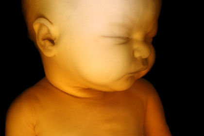
The first three months of embryonic development are known as the first trimester, that is, the first three-month period of growth. At the end of the first trimester, the embryo looks like an adult, with all major organs having been formed. It is about 3 inches (7.5 centimeters) long. Still, an embryo born during this period trimester will not survive. Additional time in the mother's womb is needed to permit further development of the organs.
At the beginning of the second trimester, the growing organism is no longer called an embryo, but a fetus. Fetal development continues through the second and third trimesters until it is ready for birth at the end of the ninth month.
Embryo diagnosis
A number of techniques have been developed to study the development of the embryo. These techniques can be used to determine the presence of problems in the growing embryo.
An ultrasound diagnosis can be performed at any time during pregnancy. Ultrasound diagnosis is a type of technology that uses high-pitched sounds that cannot be heard by the human ear. The sound is bounced off of the embryo and the echoes received are used to identify embryonic size. The technique is similar to the one used by submarines to locate underwater structures. By 18 weeks of pregnancy, ultrasound technology can detect structural abnormalities such as spina bifida (various defects of the spine), hydrocephaly (water on the brain), anencephaly (no brain), heart and kidney defects, and harelip (in which the upper lip is divided into two or more parts).
Embryonic Transfer
Imagine a baby with two mothers! At one time, that idea may have seemed absurd. Today, the practice is common. It is accomplished by a procedure known as embryonic transfer. Embryonic transfer is carried out by removing the eggs from one female and transferring them into the body of another female. The embryos have, in effect, two mothers: the one that provided the egg necessary for fertilization and the one that provided the uterus during pregnancy.
Embryonic transfer has been widely used among animal breeders to increase the number of offspring from a valuable cow, sheep, or horse. Some endangered species have benefitted from zoo breeding programs that use embryonic transfer. In humans, embryonic transfer is sometimes used as part of a fertility program. Egg donation or the use of a surrogate uterus offers hope to infertile women who have healthy eggs but lack either normal ovaries or a normal uterus.
The technique used in embryonic transfer is typified in the procedure used with domestic animals. A prize female is stimulated with hormones (organic chemicals) to produce many eggs. These eggs are then fertilized, either through normal breeding or artificial insemination, with the sperm of a champion male. Next the embryos are flushed from the uterus with a saline (salt-water) solution. Scientists use a microscope to search for the tiny clump of cells that signify an embryo at this stage. Once found, the embryos are ready for transfer. They can also be frozen for future thawing and use, if desired. When the embryos are implanted, a syringelike device delivers them into the uterus of the foster mother. If multiple embryos exist, multiple foster mothers are needed.
Breeders can typically produce six calves from one embryonic transfer. In this manner, a single prized cow can produce many calves each year. With proper training and equipment, embryonic transfer can be mastered by cattle farmers themselves.
A similar procedure is used in humans when a woman who is not able to produce eggs wishes to have a baby. Another woman is found to serve as an egg donor. The egg donor may be a close relative or may be anonymous, just as the men who donate to sperm banks are anonymous.
Several donor eggs are retrieved through a minor operation. The egg from the donor and the sperm from the male are combined in the lab in a procedure known as in vitro fertilization. The fertilized egg is then implanted in the infertile woman's uterus for a normal pregnancy and birth. Three months of hormone treatment are needed to establish the pregnancy. After that, the hormones produced normally by the woman are enough to maintain the pregnancy. Nine months later, the infertile woman gives birth to a baby to whom she bears no genetic relationship. Although much less common than in vitro fertilization, embryonic transfer offers couples a higher success rate.
Chorionic villus sampling (CVS) is the most sophisticated modern technique used to assess possible inherited, genetic defects. This test is usually performed between the sixth and eighth week of embryonic development. During the test, a narrow tube is passed through the vagina or the abdomen, and a sample of the chorionic villi is removed while the physician views the baby via an ultrasound. Chorionic villi are small hairlike projections on the covering of the embryonic sac. They are rich in both embryonic and maternal blood cells. By studying these embryonic cells, genetic counselors can determine whether the baby will have any of several defects, including Down syndrome (characterized by mental retardation, short stature, and a broadened face), cystic fibrosis (which affects the digestive and respiratory systems), and the blood diseases hemophilia, sickle-cell anemia, and thalassemia. It can also show the baby's gender.
[ See also Fertilization ]
I'd just like to express my thank you's the creator(s) of this article. I'am a College student and just recently in my Biology class, I have been assigned to report about the human embryonic development. This page has helped me a lot.
Thank you very much.
i would like to thank the author as well.
because i am doing a school project and with this website i wont have to go to anyother website because this has everything i need. also i would love to learn more about this, because your website has made this stuff very interesting(:
Destanyy(:
Can a live human embryo be successfully removed from one women and be successfully implanted alive in another woman? Has it been done? How often and with what risks and success rates? How about foetuses? Will such procedures become routine in the near future?
If so then passionate pro lifers should be willing to give birth to embryos of women who would otherwise choose to abort them.
Footnote: Thomas Aquinas and St. Augustine are generally accepted as the two greatest theologians of the Catholic Church. Both have stated life does not quicken till the immortal soul is implanted in the embryo which does not happen till the third month of pregnancy. Abortion before then would not be the murder of a human being
But I have a question, here you are saying that the "nerves, muscle, and connective tissues emerge around the primitive bone formations"
I remember hearing first the muscles/flesh forms then the bones, no? Is primitive bone formations considered bones?