Eye
The eye is the organ of sight (vision) in humans and animals. The eye works by transforming light waves into visual images. Eighty percent of all information received by the human brain comes from the eyes. These organs are almost spherical in shape and are housed in the eye (orbital) sockets in the skull.
Sight begins when light waves enter the eye through the cornea (the transparent layer at the front of the eye), pass through the pupil (the
Words to Know
Aqueous humor: Clear liquid filling the small cavities between the cornea and the iris and between the iris and the lens.
Astigmatism: Vision disorder caused by an uneven curvature in the cornea (sometimes the lens), resulting in indistinct or slightly out-of-focus images.
Cataract: A clouding of the lens of the eye.
Choroid: Delicate membrane between the sclera and the retina.
Cones: Light-sensitive nerve cells of the retina that function chiefly in bright light and are sensitive to color.
Cornea: Protective lens covering the iris.
Farsightedness: Vision disorder caused by an eyeball that is too short or a lens that is too weak; objects far away are seen easily while those up close appear blurry.
Glaucoma: Serious vision disorder caused by a buildup of aqueous humor, resulting in pressure against the retina.
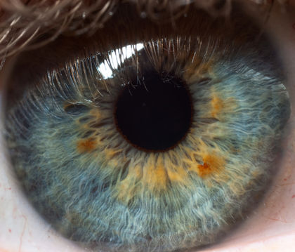
Iris: Colored portion around the pupil that regulates the amount of light entering the eye.
Lacrimal gland: Tear-producing gland that lies immediately above each eyeball at the outer corner of the eye socket.
Nearsightedness: Vision disorder caused by an eyeball that is too long or a lens that is too strong; objects up close are seen easily while those far away appear blurry.
Pupil: Adjustable opening in the center of the iris through which light enters the eye.
Retina: Photosensitive lining inside the eye.
Rods: Light-sensitive nerve cells of the retina that function chiefly in dim light.
Sclera: Tough, fibrous outer covering (the "white") of the eyeball.
Vitreous humor: Clear, gel-like substance inside the large cavity in back of the lens (the center of the eyeball).
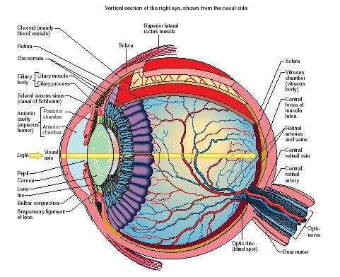
opening in the center of the colored portion of the eye, called the iris), then through a clear lens behind the iris. The lens focuses light onto the retina, which functions like the film in a camera. Nerve cells in retinas, called rods and cones, convert light energy into electrical impulses. These impulses are then carried via the optic nerve to the brain where they are interpreted as images.
The human eyeball is about 0.9 inch (2.3 centimeters) in diameter and is not perfectly round, being slightly flattened in the front and back. The eye consists of three layers: the sclera (pronounced SKLIR-a), the choroid (pronounced KOR-oid), and the retina.
Sclera
The sclera, the outer fibrous layer, encases and protects the eyeball. The visible portion of the sclera is seen as the "white" of the eye. When that portion is irritated, the small blood vessels contained in the layer enlarge, producing a "bloodshot eye." In the center of the visible portion of the sclera is the cornea, which projects slightly forward. A delicate membrane, the conjunctiva, covers the cornea and visible portion of the sclera.
Choroid
The choroid is a thin membrane lying underneath the sclera. It is composed of a dense pigment and numerous blood vessels that nourish the internal tissues of the eye. At the front end of the choroid is the ciliary body. Running like a ring around the visible portion of the eye, the ciliary body connects the choroid with the iris. The ciliary body contains muscles that are connected by ligaments to the lens behind the iris. The iris is the visible portion of the choroid. It gives the eye its color, which varies depending on the amount of pigment present in the choroid. Dense pigment makes the iris brown, while little pigment makes the iris blue. If there is no pigment the iris is pink, as in the eye of a white rabbit. In bright light, muscles in the iris constrict the pupil, reducing the amount of light entering the eye. Conversely, the pupil dilates (enlarges) in dim light, increasing the amount of light entering. Extreme fear, head injuries, and certain drugs can also dilate the pupil.
Lens
The lens is a crystal-clear, flexible body that is biconvex (curving outward on both surfaces). The entire surface of the lens is smooth and shiny, contains no blood vessels, and is encased in an elastic membrane. The lens sits behind the iris and focuses light on the retina. In addition to holding the lens in place, the muscles of the ciliary body contract and relax, causing the lens to either fatten or become thin. As the shape of the lens changes, so does its focus.
Retina
The retina is the innermost layer of the eye. The retina is thin, delicate, sensory tissue composed of layers of light-sensitive nerve cells. The retina begins at the ciliary body (not at the front of the eye) and encircles the entire interior portion of the eye. Rods and cones, nerve cells of the retina, convert light first to chemical energy and then electrical energy. Rods function chiefly in dim light, allowing limited night vision: it
is with rods that we see the stars. Rods cannot detect color, but they are the first cells to detect movement. Cones function best in bright light and are sensitive to color. In each eye there are about 126 million rods and 6 million cones.
Fluids of the eye
Between the cornea and the iris and between the iris and the lens are two small cavities. These cavities are filled with a clear watery fluid known as aqueous humor. This fluid aids good vision by helping maintain eye shape, providing support for the internal structures, supplying nutrients to the lens and cornea, and disposing of the eyes' cellular waste.
The large cavity in back of the lens (the center of the eyeball) is filled with a clear gel-like substance called vitreous humor. Light passing through the lens on its way to the retina passes through the vitreous humor. The vitreous humor is 99 percent water and contains no cells. It helps to maintain the shape of the eye and support its internal components.
Other structures of the eye
Tears are produced by the lacrimal gland, which lies immediately above each eyeball at the outer corner of the eye socket. Tears flow through ducts from this gland to the area beneath the upper eyelid. Blinking spreads the tears across the cornea's outside surface, keeping it moist and clean. Tear fluid then either evaporates or drains from the inner corner of the eye into the nasal cavity.
Eyelashes, eyelids, and eyebrows all help to protect the eye from dust and dirt. Extending from the eye socket to the eyeball are six small muscles that contract and relax, allowing the eye to move in various directions.
Vision disorders
Farsightedness and nearsightedness are common vision disorders. They occur because of a defect in the shape of the eyeball or in the refractive power (ability to bend light rays) of the lens. In these cases, the image the eye perceives is distorted because the parallel rays of light that enter the eye do not fall perfectly on a tiny hollow (called the fovea) in the retina at the back of the eye. However, corrective eyeglasses can easily overcome these disorders.
With farsightedness, objects far away are seen easily while those up close appear blurry. The cause may be that the eyeball is too short or the lens is too weak.
With nearsightedness, objects up close are seen easily while those far away appear blurry. The cause may be that the eyeball is too long or the lens is too strong.
Astigmatism, another common vision disorder, can occur in combination with farsightedness or nearsightedness. Individuals with astigmatism see indistinct or slightly out-of-focus images. The condition is brought about by an uneven curvature in the cornea (sometimes the lens). As a result, some light rays entering the eye focus on the fovea while others focus in front or behind it. Like farsightedness and nearsightedness, astigmatism can be corrected with eyeglasses or contact lenses.
A cataract is the clouding of the lens, which alters the amount of light entering the eye. The most common cataracts are senile cataracts, a result of aging that occurs in almost all people over 65 years old. These cataracts grow slowly over months or years, cause no pain, usually affect both eyes, and gradually reduce vision. If not treated, they eventually cause blindness. Clear vision can be restored by a relatively simple surgical procedure in which the entire lens is removed and an artificial lens is implanted.
Glaucoma is a serious vision disorder caused by a buildup of aqueous humor, which is prevented for some reason from properly draining. The excessive amount of fluid causes pressure against the retina, affecting vision. Long-term diseases like diabetes or a malfunctioning thyroid gland can bring about glaucoma. If left untreated, glaucoma will result in permanent blindness. The condition can be controlled with drugs that either increase the outflow of aqueous humor or decrease its production.
[ See also Radial keratotomy ]
this is zari. i have problem that when i do work on computer, using projector, multimedia, microscope or long time studies, it causes terrible headache and pain in my eyes especially right eye. i dont have any specific problem of sight still i use specs. but what really i should do i dont know.
kindly help me what is the bassic problem of my eyes and what should i do.
thank you.
Now that im older I hate it! I have one brown eye one black, a white scan inside my eye, and i have a HUGE scar on my eye lid.
Is there anything that can be done?
I had surgery in my left eye, and they took out the lens and did not put in a new one, the doctor told me (after he took out my lens) that he didnt see a point to put it back it. ( no idea why they took it out in the first place) Is there any new developments out there that I can take advantage off?
Lynda
thank u so much